
IN BRIEF
EAGLE analyzes noncontrast chest CT scans, often ordered for other reasons, for signs of esophageal cancer.
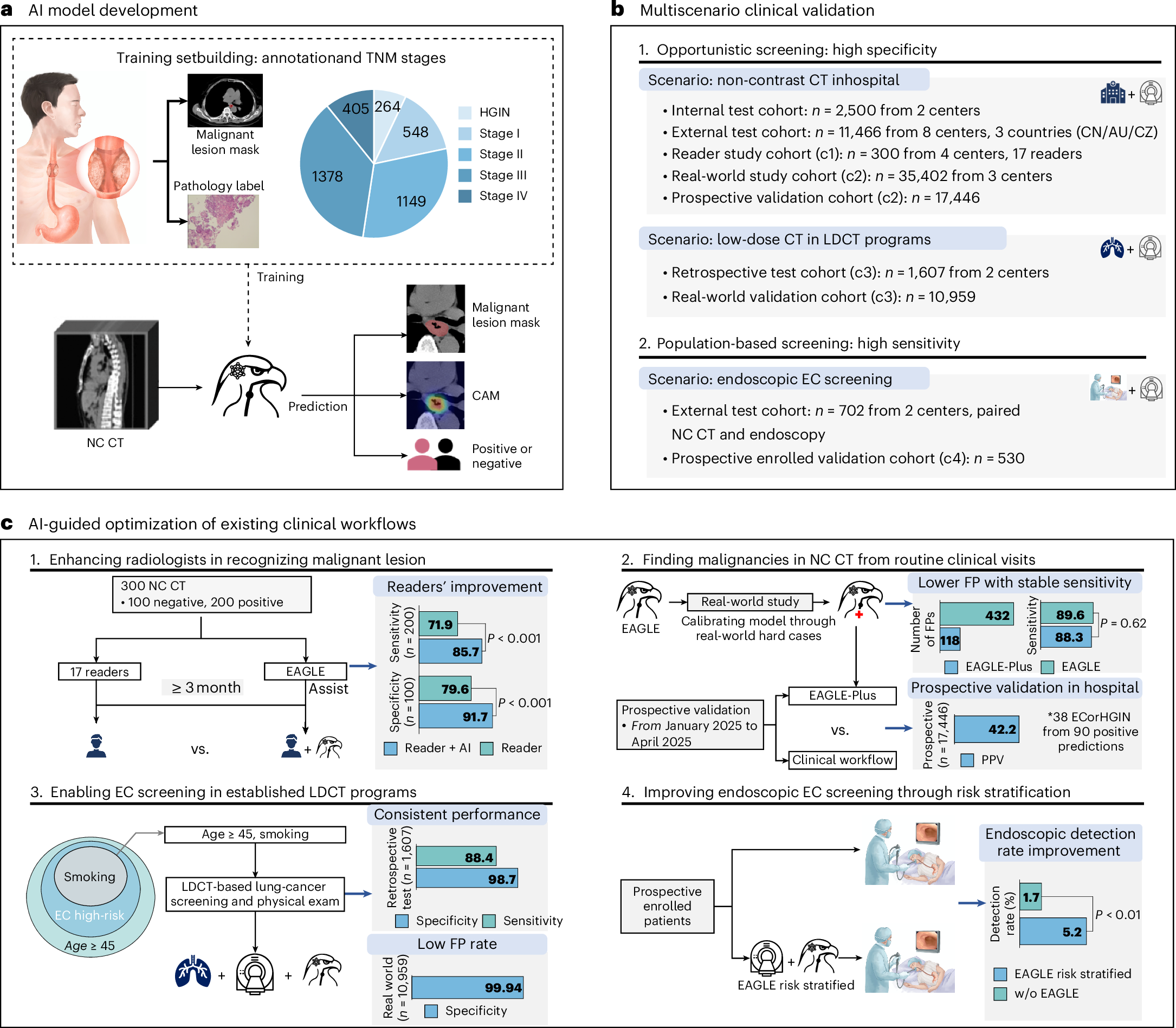
In 11,466 external scans it showed 90.0% sensitivity for cancer but 52.5% for high-grade precancerous lesions, at 98.5% specificity.
In 17,446 prospective hospital scans, 90 people were flagged and 38 confirmed. One additional cancer was diagnosed after an AI-initiated endoscopy.
WHY IT MATTERS
Esophageal cancer is frequently discovered late. CT scans already taken for thoracic conditions may contain information not routinely evaluated for subtle esophageal disease.
Automated review could highlight suspicious findings without requiring a new initial radiation exposure. But an alert is not a diagnosis, and the patient pathway after an alert determines whether screening is useful.
WHAT IS NOW POSSIBLE
Before: noncontrast CT was not routinely practical for subtle esophageal screening. Now: a model has demonstrated multicenter detection and prospective real-world alerts on 17,446 existing scans, with limited documented additional diagnosis.
WHAT MADE IT POSSIBLE
The team trained EAGLE on scans from 6,813 people at two centers, using imaging annotations informed by endoscopy and pathology.
The model returns risk classification and lesion maps. A calibrated version, EAGLE-Plus, is designed to reduce false alarms in ordinary clinical populations.
THE STORY
A chest CT scan obtained for a lung problem may also contain signs of an esophageal cancer that was not the original purpose of the examination. Researchers have developed a model to examine those noncontrast images, long thought poorly suited to identifying small lesions in the collapsed esophagus. EAGLE, described in Nature Medicine on 22 September 2026, was evaluated across hospitals and then prospectively applied to 17,446 patients.
The system was trained using 6,813 individuals from two centers. By combining CT findings with endoscopic and pathology information, the researchers produced labels for suspicious tissue even when the esophagus was hard to distinguish. Across eight external cohorts in China, the Czech Republic and Australia, involving 11,466 people, the model reached 90.0% sensitivity for esophageal cancers but only 52.5% for high-grade precancerous lesions, at 98.5% specificity. For stage I tumors, sensitivity was approximately 60.1%. The contrast shows that performance for very early disease is not the same as performance for cancers overall.
The strongest real-world test took place at SIPD, where the calibrated EAGLE-Plus system processed 17,446 consecutive eligible CT scans between January and April 2025, with follow-up of positive predictions through July 2026. It flagged 90 patients and 38 were confirmed to have an esophageal cancer or high-grade precancerous lesion, a positive predictive value of 42.2%. Across the enrolled population, 41 cancers and six high-grade precursor lesions were eventually confirmed. That does not mean all 38 flagged findings were newly discovered by AI.
Forty-eight of the 90 flagged individuals had already been handled through the standard care pathway. Of the remaining 42, a multidisciplinary group referred 16 for endoscopy, but only three completed the procedure. One had a confirmed cancer and two had negative results; four of the thirteen people who did not complete the procedure were lost to follow-up. The one confirmed cancer is the documented additional diagnosis arising from this AI-triggered endoscopy pathway, not 38 new diagnoses caused solely by the algorithm.
The researchers also tested lower-dose CT scans and strategies for deciding who merits endoscopy. Calibration across 35,402 real-world patients reduced false positives by 72.7% relative to the original algorithm, without an apparent significant sensitivity loss in the studied comparison. These findings show genuine technical progress. They do not establish that deployment reduces esophageal-cancer mortality or that the extra endoscopies and false alarms are worthwhile in every healthcare setting.
WHAT WE KNOW
External testing across eight centers found 90% cancer sensitivity and 52.5% high-grade precancer sensitivity at 98.5% specificity. [1]
In 17,446 prospectively processed scans, 90 patients were flagged and 38 confirmed, yielding 42.2% positive predictive value.
One additional cancer was documented among those undergoing AI-triggered endoscopy beyond standard care.
WHAT WE DON'T KNOW
No reduction in cancer death or complications from the model has been demonstrated.
Many people with additional alerts did not complete endoscopy, limiting verification of the real clinical pathway.
Sensitivity for high-grade precancer and very early cancer was substantially below overall cancer sensitivity.
WHAT HAPPENS NEXT
Evaluate AI-assisted referral pathways in comparative studies focused on useful early diagnoses and patient outcomes.
Validate the approach in more regions and low-dose scan types.
Assess how many extra invasive procedures and false alerts are acceptable.
SOURCE TRACE
External testing: 90% cancer sensitivity, 52.5% precancer sensitivity, 98.5% specificity.
Among 17,446 people, 90 alerts and 38 confirmed cases (PPV 42.2%).
Among 42 additional alerts, three completed endoscopy and one cancer was confirmed.
Calibration using 35,402 people reduced false positives by 72.7%.
SOURCES
- Large-scale esophageal cancer screening through noncontrast computed tomography and artificial intelligenceOpen source
- Large-scale esophageal cancer screening through noncontrast computed tomography and artificial intelligenceOpen source
CURRENT EVIDENCE
PEER-REVIEWED
REAL WORLD PROSPECTIVE CT TRIAGE / NO PATIENT SURVIVAL OUTCOME VALIDATION
WHAT WOULD CHANGE THIS?
- Comparative clinical pathways could establish how many diagnoses are genuinely brought forward.
- More complete endoscopic follow-up would strengthen false-positive estimates.
- Studies of late-stage cancer and mortality could establish whether real health outcomes improve.