
IN BRIEF
CFM combines a motorized microconfiner, distance feedback and microscopy to measure cellular traction forces on deformable gels.
When compressed, cells first show a rapid mechanical stress increase and then often a slower contractile response.
Membrane blebs may partially relieve pressure as stress stops rising or declines.
WHY IT MATTERS
Immune cells, cancer cells and developing tissues frequently move through narrow spaces. Understanding how cells handle compression could help explain migration and invasion mechanisms.
Flat-surface experiments miss some of that behavior. CFM combines dynamic control of the available space with measurements of cellular forces at the same time.
WHAT IS NOW POSSIBLE
Before: researchers had to compromise between controlled confinement, imaging and force measurement. Now: the integrated setup records a biphasic stress response and bleb-associated relaxation.
WHAT MADE IT POSSIBLE
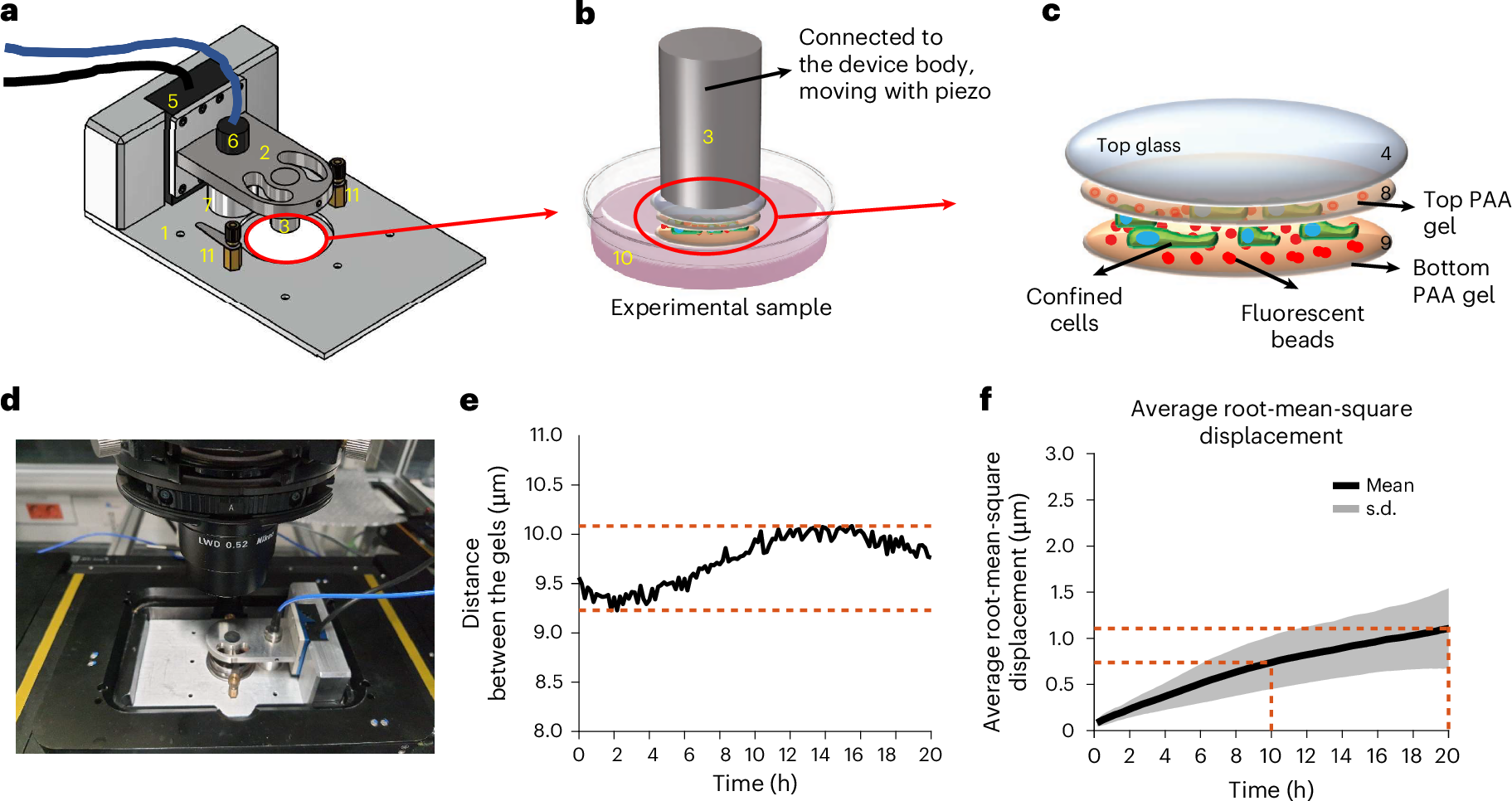
A piezoelectric stage adjusts the separation of soft gel surfaces while a capacitive sensor monitors their displacement.
Embedded fluorescent beads move with the gels, allowing reconstruction of lateral and normal traction stresses.
THE STORY
A cell moving through living tissue rarely occupies an open space. It must squeeze between obstacles, deform its nucleus and respond to physical forces. Researchers have built a system that reveals what happens while the surrounding gap changes. Called confinement force microscopy, or CFM, the method was published in Nature Methods on 14 September 2026 with experiments on living cells.
The apparatus combines a piezoelectric stage and a capacitive displacement sensor to control the gap between two soft substrates. The researchers can change compression during live imaging. Fluorescent particles embedded in the gels move as cells push and pull, and their displacement is used to reconstruct three-dimensional traction stresses, both parallel and perpendicular to the substrate.
The team measured platform stability for as long as twenty hours. Across tests, average drift remained around a micrometer over the full period, enabling observation beyond the first seconds of compression. The instrument was used with several cell types, including HeLa cells, C2C12 cells and neutrophils, although some detailed experiments involved smaller cell subsets.
A recurring two-stage pattern appeared. Compression first created a fast rise in stress consistent with passive deformation of the cell and nucleus. This was often followed by a slower increase linked to actomyosin contractility and intracellular pressure. Many cells then formed rounded membrane protrusions called blebs. During their expansion, traction stresses frequently stabilized or declined, suggesting partial relief of internal pressure.
The researchers also inhibited ROCK and myosin II to examine mechanisms underlying the second stage. Both interventions reduced stress generation but changed bleb formation differently. The experiments help distinguish passive physical loading from an active cellular response. They remain cell-culture findings, not direct measurements of tumor invasion in patients or proof of a new therapy. CFM offers a better way to investigate those future questions.
WHAT WE KNOW
The platform controls confinement dynamically while measuring lateral and normal cellular stresses. [1]
It maintained useful displacement stability during experiments lasting up to twenty hours.
Cells frequently showed an early passive peak, later active stress and membrane blebs.
WHAT WE DON'T KNOW
Gel confinement cannot reproduce every physical feature of living tissue.
The exact benefit or harm of bleb formation under physiological conditions is unresolved.
No clinical diagnostic or treatment advantage has been demonstrated.
WHAT HAPPENS NEXT
Repeat measurements in more realistic tissue models.
Compare mechanically different immune, cancer and normal cells.
Test whether the measured stress signatures explain migration in biological tissues.
SOURCE TRACE
CFM controls compression and measures lateral and normal cell stresses.
Mean gap drift was about one micrometer over 20 hours.
Passive stress was followed by active contraction and blebs with possible relaxation.
SOURCES
- CFM: confinement force microscopy—a dynamic, precise and stable microconfiner for traction force microscopy in spatial confinementOpen source
- CFM: confinement force microscopy—a dynamic, precise and stable microconfinerOpen source
CURRENT EVIDENCE
PEER-REVIEWED
LIVING CELL LAB EXPERIMENT / NOT CLINICAL
WHAT WOULD CHANGE THIS?
- Independent replication could establish response patterns across more cell types.
- More realistic three-dimensional environments could strengthen biological relevance.
- In-vivo experiments could link the measurements to tissue behavior.